Fluorescence Microscopy
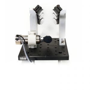
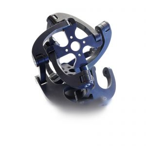
Filter Cube Switcher
 Highlights
- Speed 10× vs state of the art
- Low vibration mechanical configuration
- Dichroic and emission filter switching
Background
Traditional filter cube turrets used in modern fluorescence microscopes perform exceptionally well at processing excitation and emission light with filter sets precisely designed for each individual fluorescent label used in a given multi-stained specimen. Live cells offer unique insight into biological processes but also present a challenging observation target, not the least due to the transient nature of the subject of the study. The filter exchange speed of a cube turret limits peak optical performance of single-band filters to experiments with dynamics on the order of seconds, faster examination is only possible with multi-band dichroic filters and multi-band emission filters, single-band emission filters loaded in a high-speed filter wheel, or an image-splitting device.
Approach
 High-performance LED or laser light sources deliver light of excellent spectral and beam quality and switch between individual wavelengths in tens of microseconds. They also enable the simplification of the traditional cube turret by eliminating the excitation filters, a concept exploited in dichroic and emission filter changer design aimed at achieving speeds associated with high-speed filter wheels. While convenient, fluorescence filter cubes, especially the precision metal type, are heavy and therefore difficult to move quickly. In this project, exchange speed is maximized by loading the dichroic and emission filters onto a pair of light-weight carriages that are actuated simultaneously in opposite direction. While increasing speed by reducing the inertia of the moving parts, this approach also reduces vibration by cancelling out reaction forces to the acceleration and the deceleration of the counterpoised loads. A special dichroic filter attachment mechanism was devised that securely retains the filters without compromising its surface flatness, making the system suitable for TIRF microscopy. The moving parts are assembled in preloaded configuration to achieve high repeatability of the filter orientation required for photostimulation experiments.
Specifications
| Filter Count | 5 pairs |
| Adjacent Filter Switch | 30ms |
| Dichroic Filter Size | 25mm × 36mm – 26mm × 38mm | 1mm or 2mm thick |
| Emission Filter Size | ø25mm | ø1″ | 3.5mm or 5mm thick |
| Excitation Filters | External |
| Interface | Digital trigger | RS-232 |
High Speed Filter Cube Change
Adjacent dichroic and emission filter exchange in 30ms.
DIC and Fluorescence Microscopy
Zebrafish embryo bloodflow acquired in DIC and fluorescence modalities. (video credit: Simon Watkins, Center for Biologic Imaging, University of Pittsburgh)
DIC and Fluorescence Microscopy
Zebrafish embryo heartbeat acquired in DIC and fluorescence modalities. (video credit: Simon Watkins, Center for Biologic Imaging, University of Pittsburgh)