Imaging Path Switching in 3ms
 Highlights
- Emission filter switching in 3ms, standard ø25mm size
- Compatibility with light carrying embedded spatial information
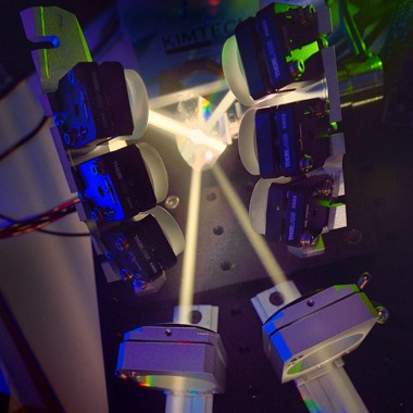
- Single input port, single output port, four intermediate paths
Background
Examination of fast biological processes using fluorescence microscopes has become possible with the advent of scientific imaging cameras equipped with sensitive EMCCD or sCMOS detectors and fast data readouts. Rapid excitation wavelength switching on the other end of the light path is facilitated with LED and laser light sources but inertia associated with physical filter exchange in filter wheels limits emission wavelength switching to about 23ms.
State of the art
Experiments that fully exploit the throughput of modern scientific cameras must rely on emission wavelength discrimination using multi-band filters. The design of these filters is more complex and their performance is inferior when compared to single band filters manufactured using the same technology. Alternatively, image-splitting devices that allow simultaneous multi-color imaging are commercially available, but suffer in varying degrees from bleed through between adjacent channels. Multicolor fluorescence microscopy imaging using single-band emission filters at millisecond time scale could provide new insights into highly dynamic cellular processes.
Approach
Physical filter switching is fundamentally limited by the inertia of the moving mass and design tradeoffs lead to impractical solutions with switching times faster than about 20ms. In order to move beyond this threshold, instead of physically repositioning an array of optical elements, light can be directed by a fast mirror to a series of individual optical paths and then recombined to a common light path using another fast mirror. The obvious disadvantage of this approach is the use of two high-speed mirrors. A better arrangement, such as the one developed in this project, employs the same mirror that is used to direct the light along different light paths also for descanning. This novel solution reduces the overall component cost and provides a basis for a simpler, more reliable and compact design. The optical design of a microscope imputes challenging constraints on the parameters and layout of the components of the path switching instrument. The space between the output port of the microscope and the image plane is not sufficient for the implementation of the switcher but it can be extended with a finite conjugate relay. It is important to minimize the negative impact of introducing additional lenses and mirrors on the quality of the light coming out of the microscope. The parameters of the relay lenses were defined such that optical aberrations are minimized and no light falloff occurs even with high numerical aperture objectives and precision ultra-flat mirrors with high-reflectance coatings are used throughout the assembly.
Specifications
| Number of Alternative Paths | 4 |
| Adjacent Patch Switch | 3ms |
| Filter Size | ø25mm | ø1″ |
| Efficiency | 85% |
| Field Size | ø18mm |

